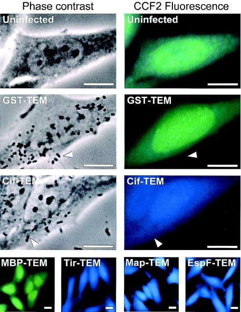
FIG. 2.
Demonstration of the translocation of EPEC effector proteins into live HeLa cells by using TEM-1 fusions and fluorescence microscopy. HeLa cells were infected with wild-type EPEC strains expressing different TEM-1 fusion proteins. After infection, HeLa cells were washed and loaded with CCF2/AM. β-Lactamase activity in HeLa cells is revealed by the blue fluorescence emitted by the cleaved CCF2 product, whereas uncleaved CCF2 emits a green fluorescence. No detectable fluorescence arises from adherent bacteria (indicated by arrowheads). Bars, 10 μM.

