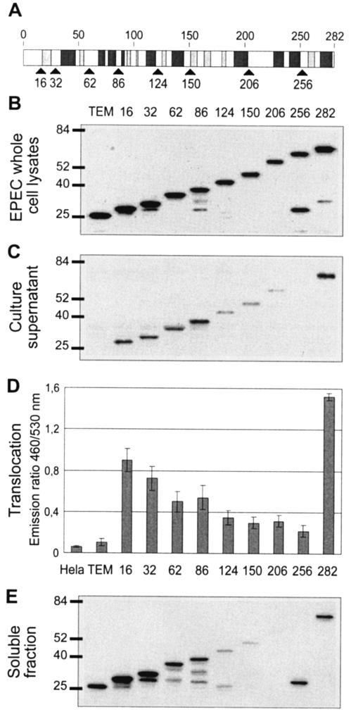
FIG. 4.
Identification of the minimal N-terminal domain of Cif that can mediate translocation into eukaryotic cells. (A) Schematic representation of the predicted secondary structure of Cif. Light and dark gray boxes represent β-sheets and α-helices, respectively. Black arrows indicate the different sites of fusion to TEM-1. (B) Expression of Cif1-X-TEM fusions in EPEC. EPEC whole-cell lysates were subjected to Western blot analysis with anti-TEM-1 antibody. Molecular mass markers (in kilodaltons) are indicated to the left. (C) Secretion of the produced Cif1-X-TEM fusions. Culture supernatants from E22 Δcif expressing TEM-1 alone and Cif1-X-TEM fusions were subjected to Western blot analysis with anti-TEM-1 antibody. (D) Translocation of Cif1-X-TEM fusions in HeLa cells. The presented data are averages of triplicate values of the results from three experiments. Numbers indicate the different sites of Cif fused to TEM-1. (E) Solubility analysis of the produced Cif1-X-TEM fusions. EPEC cells were lysed, and the soluble fraction was obtained after the removal of insoluble proteins, cell debris, and unbroken cells by centrifugation.

