. 2016 May 16;5:e13964. doi: 10.7554/eLife.13964

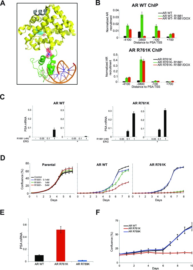
Figure 4. R761 methylation regulates AR recruitment, transcription, and proliferation.

(A) Model of AR LBD (PDB: 2AO6; yellow) and AR DBD (PDB: 1R4I; green) interactions (see Materials and methods). A modeled di-methylated R761 is shown (red). Grey ribbon: TIF2 coactivator peptide. Cyan spheres: R1881. DNA is shown as orange/blue sticks. (B) AR ChIP qPCR for regions of the PSA gene as in Figure 2D from RWPE-1 cells expressing wild-type (WT, left) AR or AR R761K (right). DOX: ERG expression; R1881 is 1nM. Error bars represent + SEM of three biological replicates, each with three technical repeats. (C) PSA qRT-PCR in RWPE-1 cells expressing WT AR (left) or AR R761K (right). Error bars represent + SEM of three biological replicates, each with three technical repeats. (D) RWPE-1 parental cells and cells expressing either wild type AR (AR WT) or AR R761K mutant were left either untreated (control) or treated with 0.1, 1 or 5nM R1881 for 6 days and confluence measurements (see Materials and methods for description) were collected. Error bars represent + SEM of three biological replicates, each with three technical repeats. (E) PSA qRT-PCR from VCaP cells expressing the noted AR constructs, grown in androgen-depleted media (charcoal-stripped serum). (F) VCaP cell proliferation upon expression of the noted AR constructs.

DOI: http://dx.doi.org/10.7554/eLife.13964.013

Figure 4.

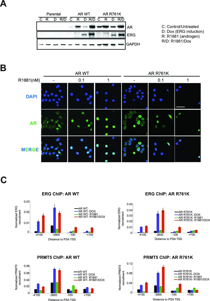
Figure 4—figure supplement 1. AR R761K mutation effects in RWPE-1.

Figure 4—figure supplement 1.

(A) Western blot of RWPE-1 parental cells and cells stably expressing AR wild type (AR WT) or AR R761K mutant. Cells were either untreated (C) or treated with 100ng/ml doxycycline (D) to induce ERG expression, in the absence or presence of 1 nM R1881 for 24 hr (R, R/D). Blots for AR, ERG, and GAPDH are shown. (B) Representative immunofluorescence images of AR from RWPE-1 cells stably expressing either AR WT or R761K mutant. Cells were either untreated (-) or treated with 0.1 or 1nM R1881 for 24 hr. Cells were also stained with DAPI to visualize nuclei. Cells were fixed and stained as described in Materials and methods. Scale bar, 50 µm. Data shown is a representative example of three biological replicates. (C) ChIP qPCR of ERG (top row) and PRMT5 (bottom row) at the PSA promoter and enhancer regions in RWPE-1 cells expressing either wild type AR (AR WT, left column) or AR R761K mutant (right column) in the absence and presence of ERG (Dox) or R1881 (R; 1nM for 24 hrs). All recruitments are normalized to the IgG control ChIP. Error bars represent + SEM of three biological replicates, each with three technical repeats.