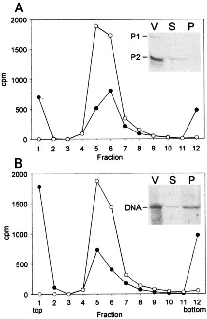
FIG. 2.
Association of l-[35S]methionine- (A) and 33P- (B) labeled PM2 virions with ER72M2 cells. The distribution of the radioactivity was analyzed by rate zonal centrifugation after a 15-min adsorption period using an MOI of 10 (black circles). A corresponding amount of labeled virus particles alone was used as a control (open circles). The radioactivity on top of the control gradient (virus only) was subtracted from the adsorption experiment to correct the effect of spontaneously dissociated phage particles. The insets show the supernatant (S) and the cell fraction (P) of an adsorption mixture (MOI, 5) and the labeled virions (V) analyzed in a tricine-polyacrylamide gel followed by autoradiography. The positions of PM2 P1 and P2 proteins and DNA are indicated.

