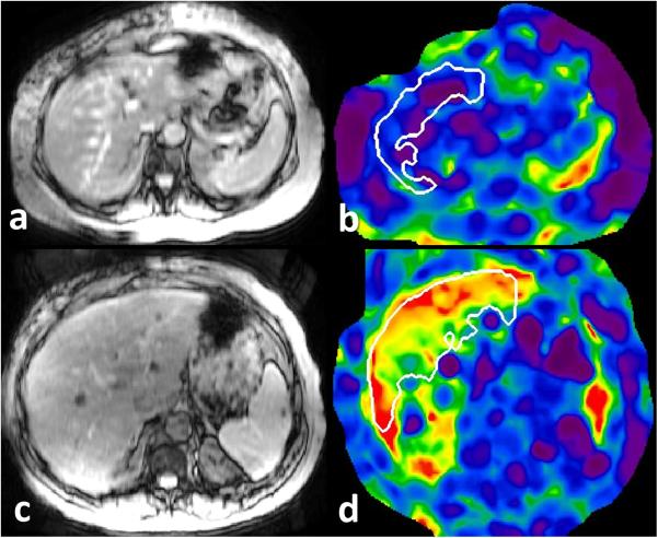
Figure 1.
MRE of liver in patients with suspected constrictive pericarditis. Top row: magnitude MRE image (a) and corresponding stiffness map (b) of liver in a 59 year old male with echo negative for CP and estimated right atrial pressure of 5mm Hg has a normal liver stiffness of 1.98kPa. Bottom row: magnitude (c) and stiffness map (d) in a 61 year old male with echo diagnostic of CP and estimated right atrial pressure of 14mmHg has elevated liver stiffness of 5.4kPa (normal cut-off value is 2.5kPa). The white outlined region represents region of interest drawn using automated algorithm avoiding liver edges and large vessels.

