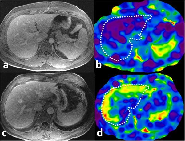
Figure 2.
MRE of liver in patients with suspected constrictive pericarditis. Top row: post gadolinium enhanced delayed T1-weighted axial image (a) and corresponding level stiffness map (b) of liver in a 26 year male with echo negative for CP and estimated right atrial pressure of 5mm Hg. The liver has normal morphology and a normal mean liver stiffness of 1.99kPa. Bottom row: post gadolinium enhanced delayed T1-weighted axial image (c) and stiffness map (d) in a 65 year old male with echo diagnostic of CP and estimated right atrial pressure of 20mmHg has elevated liver stiffness of 4.4kPa. Note the normal morphology of liver but prominent infewrior vena cava. The white dotted outline represents liver on the stiffness maps.

