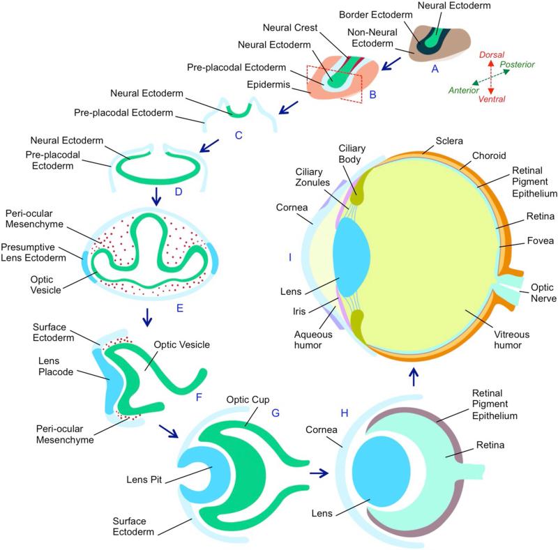
Figure 2. Eye development in vertebrates.
(A) During gastrulation, the ectoderm is divided into three distinct regions - Neural ectoderm, non-neural ectoderm and the border ectoderm region between these tissues. (B) The border ectoderm gives rise to pre-placodal ectoderm neural crest cells. Red dotted rectangle indicates a section through the embryo that is represented in (C). (C) The neural ectoderm cells comprising the neural plate fold inwards to form the neural tube. (D) A region of cells within the neural ectoderm (anterior neural plate) is specified by eye field transcription factors to form a single eye field, which by Sonic Hedgehog signaling, is partitioned into bilateral optic sulci. (E) Each of the optic sulci develops into an optic vesicle that migrates towards the non-neural ectoderm, which is specified as the surface ectoderm or pre-placodal ectoderm. (F) Interactions between the optic vesicle and the pre-placodal ectoderm results into the latter forming the lens placode. The surrounding peri-ocular mesenchyme inhibits the surface ectoderm that does not appose the optic vesicle from acquiring lens fate. (G) Subsequently, the lens placode and the optic vesicle coordinately invaginate to form the lens pit and optic cup, respectively. (H) The lens pit continues to invaginate the optic cup until it pinches off to form the lens, while the overlying surface ectoderm contributes toward the cornea. The optic cup forms the neuro-retina and the retinal pigment epithelium. (I) Subsequent development and differentiation results in the formation of a multicomponent eye. In the anterior region, the adult eye contains the outer cornea, the iris, the lens, the ciliary body and ciliary zonules, while in the posterior region, it contains the retina. The space between the cornea and the lens is occupied by aqueous humor, while that between the lens and the retina is occupied by the vitreous humor. Light is focused by the cornea and the lens on the retina. The iris responds to the intensity of the light and changes its pinhole similar to the aperture of a camera. The focusing power of the lens is mediated by the ciliary zonules, arising from the ciliary body. Photoreceptor cells within the retina convert the photon energy in light into electrical signals that are transmitted by the optic nerve to the brain where it is interpreted as an image. The retinal pigment epithelium has several functions such as light absorption, nutrient transport, and reduction of photo-oxidative stress by photoreceptor membrane renewal. The fovea is a location in the retina where there is a high concentration of cone photoreceptor cells and where visual sharpness is high.

