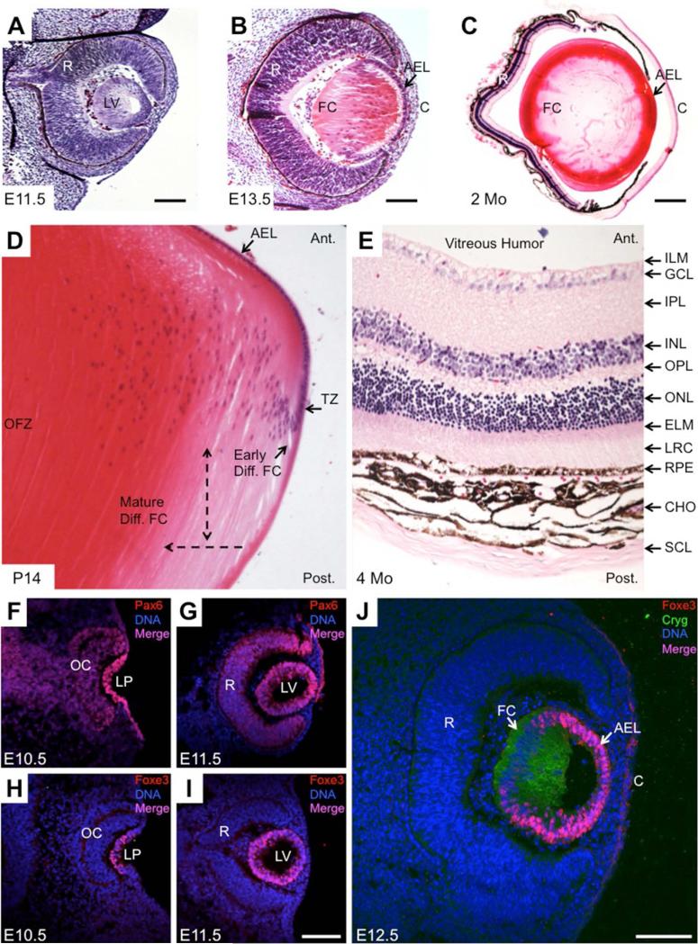
Figure 3. Phenotypic characteristics of the developing mouse eye.
Representative embryonic and postnatal mouse eye tissue stained with hematoxylin and eosin stains that bind to nucleic acid and protein rich regions in the cell, respectively, are shown in A through E. Immunofluorescence maker analyses of key genes in mouse lens development are shown in F through J. (A) At E11.5, a hollow lens vesicle is observed, in which posteriorly located cells have initiated differentiation into primary fiber cells. Also observed is the retina that is primarily composed of the retinal progenitor and ganglion cells. (B) At E13.5, the lumen of the lens vesicle is filled with elongated primary fiber cells and the epithelial cells can be seen in the anterior of the lens. The overlying surface ectoderm will form the cornea. (C) At age 2 months, an adult lens exhibits an epithelial cell layer at the anterior region and fiber cells in the posterior region. The cornea has several layers of cells and the retina is developed with a laminated structure. (D) High magnification image of a post-natal day (P) 14 mouse lens. In the transition zone, cells of the anterior epithelium exit the cell cycle and initiate differentiation of fiber cells. Single head broken arrow indicates direction of early to mature differentiating fiber cells, while double head arrow indicates direction of fiber cell elongation. Terminally differentiated mature fiber cells form a central nuclear-free region called the organelle free zone in the lens. (E) Adult mouse retina is a laminated structure comprising of eleven distinct layers of cells. The sclera originates from the neural ectoderm and protects the eye globe. (F) At E10.5, a critical regulator of eye development Pax6 exhibits expression and nuclear localization in cells of the lens pit and the optic cup. (G) At E11.5, Pax6 continues to be expressed in the lens vesicle and in the retina. (H) A lens-enriched transcription factor Foxe3 is expressed in the lens pit at E10.5. (I) At E11.5, Foxe3 is expressed in all the cells of the lens vesicle. (J) In the following stages, Foxe3 protein is restricted to the cells of the anterior epithelium of the lens. Gamma-crystallin staining is a marker for differentiated lens fiber cells. Abbreviations: LV, Lens Vesicle; R, Retina; AEL, Anterior Epithelium of the Lens; C, Cornea; FC, Fiber Cells; TZ, Transition Zone; OFZ, Organelle Free Zone; Ant., Anterior; Post., Posterior; ILM, Inner Limiting Membrane; GCL, Ganglion Cell Layer; IPL, Inner Plexiform Layer; INL, Inner Nuclear Layer; OPL, Outer Plexiform Layer; ONL, Outer Nuclear Layer; ELM, External Limiting Membrane; LRC, Layer of Rods and Cones; RPE, Retinal Pigment Epithelium; CHO, Choroid; SC, Sclera; OC, Optic Cup; LP, Lens Pit. Scale bar in A, B, I, J, is 100 μm; C is 400 μm.

