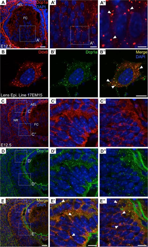
Figure 6. Processing bodies in mouse lens and retina development.
(A-A”) The Processing body (P-body) marker Dcp1a is observed to stain distinct granules in the mouse lens at embryonic day (E) 12.5. P-bodies are RNA granules that undertake mRNA storage, decay or silencing. (B-B”) A second P-body marker, Ddx6, stains distinct granules and co-localizes with Dcp1a in the mouse lens epithelial cell line 17EM15. (C-E’’) P-body markers Ddx6 and Dcp1a stain distinct granules and co-localize in the E12.5 mouse retina. Scale bar in A, E is 25 μm; in A’, B’’, E’, E’’ is 10 μm; in A’’ is 5 μm.

