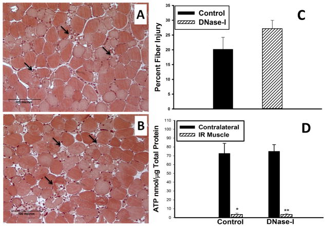
Figure 2.
Effect of DNase-I treatment on muscle fiber injury and ATP levels following acute IR. Image A and B, are representative micrographs of Mason’s trichrome stained acrylic imbedded TA muscle shows the muscle tissue morphology in cross sections following IR from control (A) and DNase-I treated (B) mice. Injured muscle fibers are evident in the field such as the one indicated by black arrow with observed edema, hemorrhage, and leukocytes infiltration. Quantitative analysis of the percent muscle fiber injury per randomized per high power field is summarized in the graph (C), and muscle ATP summarixed in the graph (D) revealed that, DNase-1 treatment did not alter skeletal muscle fiber injury or the steady state levels of ATP.

