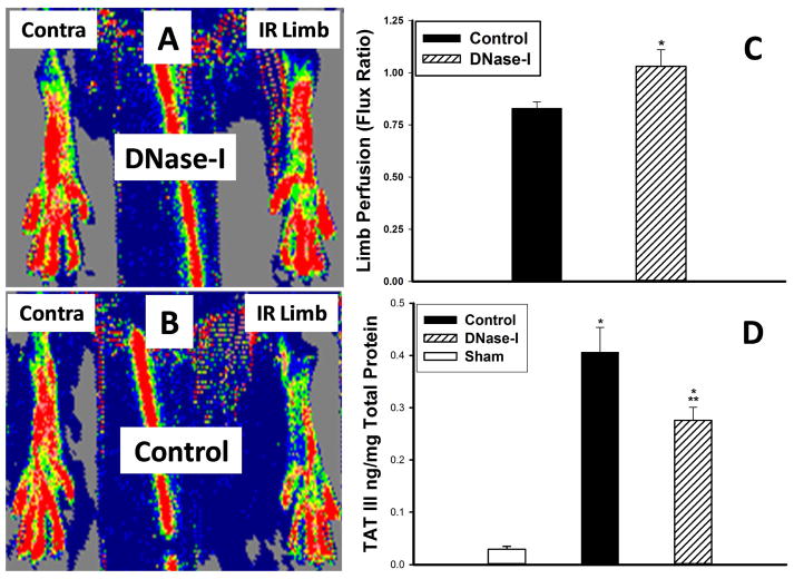
Figure 3.
Effect of DNase-I treatment on hind limb perfusion and TAT III levels in the hind limb muscle following IR. The Laser Doppler scanner generated images (A&B) demonstrates the degree of perfusion in each hind limb (red to be the highest perfusion in contrast to dark blue which represent no perfusion). The IR limb in the Pulmozyme treated mouse (Figure-2A) appeare to have similar perfusion compared to the uninjured contralateral limb while the IR limb in the untreated control mice (Figure-2B) have diminished perfusion compared to the contralateral limb. Calculating the hind limbs the scanner generated Flux ratio the hind limbs in each mouse as an index of limb perfusion revealed, significantly higher perfusion at 24 hours reperfusion in the DNase-I treated mice compared to the control group (*p=.02, C). Hind limb tissue TAT III levels significantly increased following IR in both control and the DNase-I treated group compared to sham (* p<.001). However the TAT III levels were significantly lower in the DNase-I treated mice compared to control (**p<.05). This data suggest that DNase-I treatment ameliorated tissue thrombosis which parelled higher hind limb perfusion.

