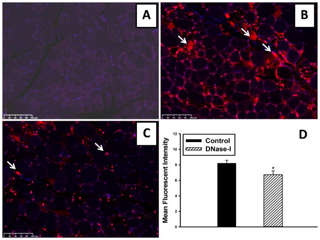
Figure 4.
Effect of DNase-I treatment on ETs detection following IR. Representative images from Cy3 labeled ETs immunostaining. ETs wer not detectable in the non injured sham muscle (A). However, there was extensive ETs detection observed in the interstitium of the injured muscle fibers as indicated by white arrows (B). DNase-1 treatment significantly reduced ETs detection in the tissue following IR (C). Mean fluorescent intensity values for DNase-I treated and untreated control mice are summarized in the graph (*p=.036, D).

