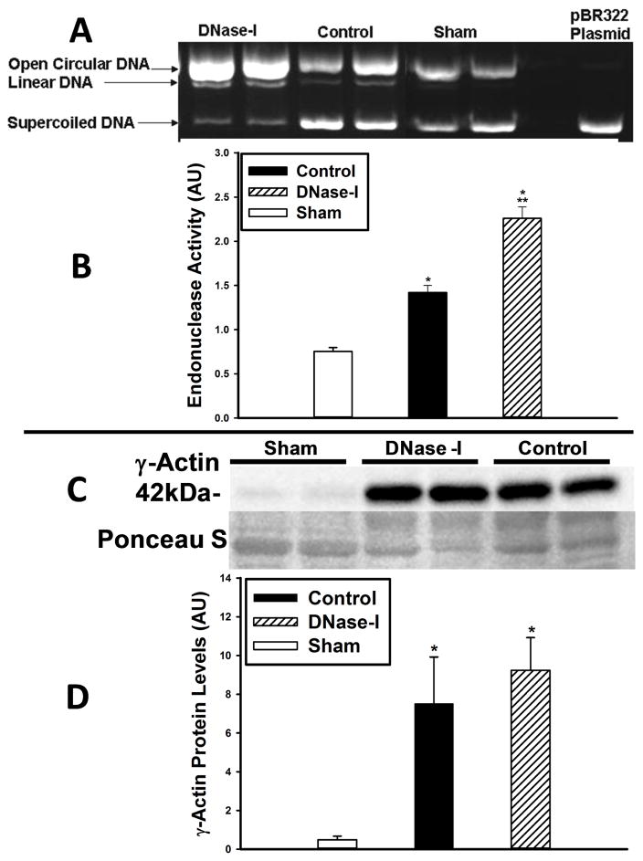
Figure 6.
Effect of DNase-I treatment of endonuclease activity and γ-Actin protein levels following IR. Endonuclease activity was evaluated with plasmid incision assay in skeletal muscle tissue protein extracts from sham, DNase-I treated and untreated control mice (A&B). The representative image (A) show the bands for the open circular and linear forms of the plasmid DNA which increased over the supercoiled DNA in the DNase-I treated and control groups compared to sham indicating increased endonuclease activity while the right lane of the gel image represent the pBR322 plasmid alone which only present a supercoiled plasmid DNA form. Quantitative analysis (B) shows that endonuclease activity was markedly elevated in both the control and DNase-I treated groups over sham levels (*P<0.01, B) with significantly higher levels detected in the DNase-I group compared to control (**P< .05, B). This data suggest that DNase-I treatment further enhanced endonuclease activity following IR. Analysis of γ-Actin protein expression by western blotting, shows an enhanced γ-Actin protein expression following IRas compared to sham (C). Qquantitative analysis of the 42kDa γ-Actin band densities normalized to the Poncaue-S bands are displayed in the summary graph (D). Data shows a significant increase γ-Actin protein in the post ischemic hind limb muscle in the DNase-I treated and control groups compared to sham (* p<.01, D). There was no difference in the γ-Actin levels between the DNase-I treated and control groups (D). This data suggest that DNase-I treatment did not alter the γ-Actin protein levels.

