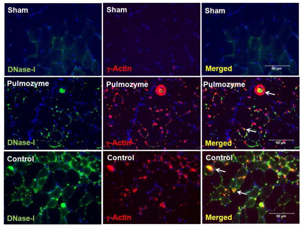
Figure 7.
Immunofluorescence localization of DNase-I with γ-Actin proteins in skeletal muscle tissue following IR with and without exogenous DNase-I treatment. Images in the panel are representative of dual immunofluorescence detection of DNase-I and γ-Actin using Alexa-488 and Cy3 fluorochrome conjugated secondary IgG respectively in skeletal muscle tissue from uninjured sham hind limb and following IR. In the sham mice (top images), there was very low level of DNase-I (top left, green) or γ-Actin (top left middle, red) and merged (tope right), detected in the tissue. However, we observed in the mice that were treated with DNase-I (Pulmozyme,(middle images) or in the control group (lower images) extensive DNase-I (Green color) and γ-Actin (Red color) staining detected at 24 hours following IR. Merged fluorescent images demonstrated that DNase-I and γ-Actin molecules colocalized within and around the injured fibers in the DNase-I treated and control groups (merged colors in yellow/Orange). The colocaliation of DNase-I suggest that DNase-I might be inhibited in the presence of γ-Actin in the injured tissue.

