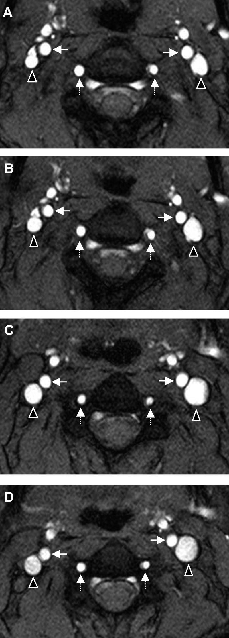
Fig. 1.
Cross-sectional magnitude images from a phase-contrast MRI sequence taken between the second and third vertebrae in one subject. The images show the left and right internal jugular veins (triangles), internal carotid arteries (solid arrows), and vertebral arteries (dashed arrows) at 0° baseline (A), −6° HDT (B), −12° HDT (C), and −18° HDT (D).

