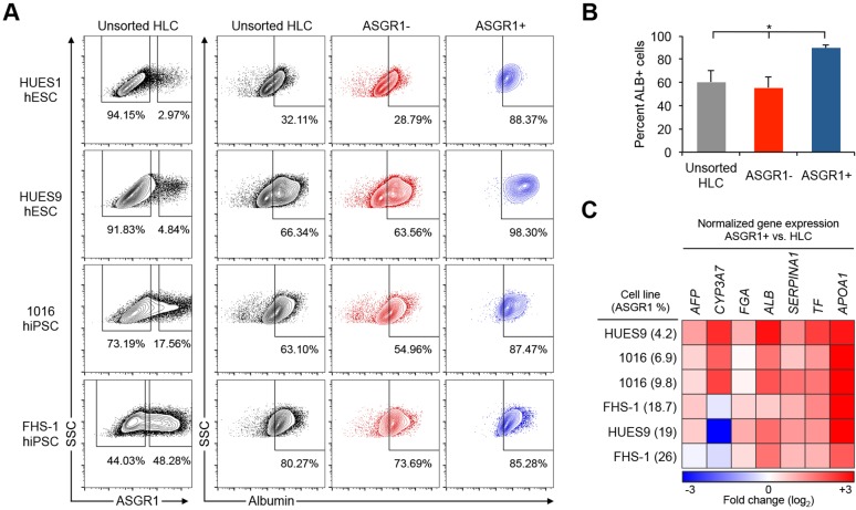
Fig. 2.
Enrichment of hepatocytes from HLC differentiation cultures by surface ASGR1 FACS. (A) Four different hPSC lines were differentiated to HLCs. The percentage of cells expressing the hepatocyte marker ALB among unsorted HLCs, surface ASGR1-negative cells, and surface ASGR1-positive cells was quantified by intracellular flow cytometry. (B) Summary of results in A showing the mean percentage of ALB-positive cells by flow cytometry among unsorted HLCs, surface ASGR1-negative cells and surface ASGR1-positive cells (n=4 differentiations). Error bars represent s.e.m. *P<0.05, Student's t-test. (C) Heatmap summarizing qRT-PCR results, showing relative expression levels in ASGR1-positive cells compared with matched unsorted HLCs.

