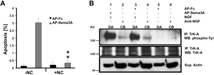
Fig. 3.
Retrograde Sema3A-induced cell death requires microtubule-based axonal transport in sympathetic neurons. (A) In compartmentalized cultures of sympathetic neurons, distal axons were exposed to either AP-Sema3A or AP-Fc, with or without nocodazole (NC). Six hours after stimulation, immunofluorescent labeling of activated caspase 3 was performed, followed by quantification of activated caspase 3+ cell bodies. The short 6 h treatment was used, even though this resulted in a lower percentage of Sema3A-mediated apoptosis, because NC alone inhibits retrograde transport of NGF-TrkA complexes, which will result in apoptosis of the neurons within 24 h. Mean±s.e.m. from three independent experiments. **P<0.01, Student's t-test. (B) In three separate SCG compartmentalized cultures (lanes 1 and 2, 3 and 4, 5 and 6), distal axons (DA) were treated with control AP-Fc (lanes 3 and 5) or AP-Sema3A (lane 1) with or without NGF. In some conditions the cell bodies (CB) were treated with anti-NGF (lanes 2, 4 and 6). After 24 h, the neurons were subjected to TrkA immunoprecipitation followed by western blotting with the indicated antibodies. Sema3A treatment of axons in the presence of NGF does not affect TrkA activation or its retrograde transport to cell bodies (lanes 1 and 2), when compared with neurons treated with AP-Fc and NGF (lanes 3 and 4). Supernatants from the immunoprecipitations were analyzed for actin as a loading control. Similar results were obtained from three independent experiments.

