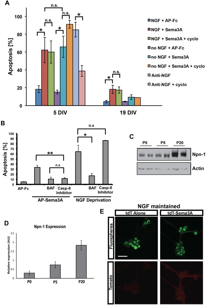
Fig. 4.
Sema3A induces apoptosis via the extrinsic pathway. (A) SCG neurons were maintained for 5 DIV or 19 DIV, at which time they are fully mature. The neurons were then deprived of NGF, deprived of NGF in the presence of cycloheximide, given Sema3A in the presence or absence of NGF, or given Sema3A in the presence of cycloheximide for 24 h. The percentage of neurons that underwent apoptosis was quantified by measuring both cellular morphology and nuclear morphology. Mean±s.e.m. from three independent experiments. (B) SCG neurons were maintained in vitro for 5 DIV and then exposed to AP-Fc, AP-Sema3A, or deprived of NGF in the presence or absence of the pan-caspase inhibitor BAF (50 µM) or caspase 8 inhibitor II (5 µM). Apoptosis was ascertained 24 h later as described for A. Mean±s.e.m. from three independent experiments, except for the AP-FC treatment (n=2). (A,B) n.s., not significant; *P<0.05, **P<0.01, Student's t-test. (C) Sympathetic neurons maintained in culture to the equivalent of P0, P5 or P20 were detergent extracted and subjected to Npn1 immunoblotting after immunoprecipitation of Npn1. Note that Npn1 expression increased by P20 relative to actin, which was used as a loading control. (D) The experiments in C were quantified from three independent cultures; mean±s.e.m. (E) Retrograde Sema3A transport studies of compartmentalized cultures of P18 SCG neurons. Note that at this age little or no tdT-Sema3A was retrogradely transported, as compared with the transport of tdT alone. Scale bar: 50 μm.

