Figure 2.
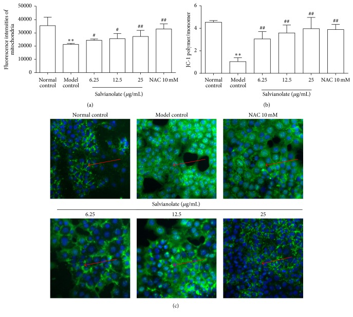
Protective effects of Salvianolate on H2O2-induced mitochondrial injury in AML-12 cells in vitro. Hepatocytes injury model was established with 0.5 mM H2O2, and then the cells were incubated with Salvianolate with concentrations of 6.25, 12.5, or 25 μg/mL or NAC with concentration of 10 mM for 24 h. (a) Semiquantification data for expressions of viable mitochondria in AML-12 cells by quantifying the fluorescence intensity of mitochondria probe with MitoTracker Green kits. (b) Semiquantification data for expression of MMP in AML-12 cells by examining the fluorescence intensity ratio of JC-1 aggregation/JC-1 monomer. (c) Expression of Cyto-C (green fluorescence marked with red arrows) in cytoplasm was revealed by the images taken by Cellomics ArrayScan VTI HCS Reader. ∗∗ p < 0.01 versus normal control; # p < 0.05 and ## p < 0.01 versus model control.

