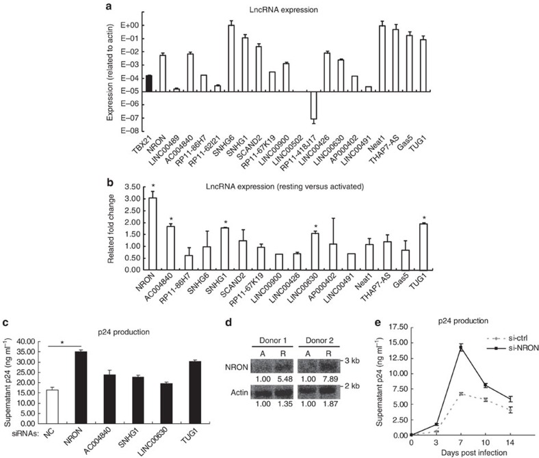
Figure 1. LncRNA NRON represses HIV-1 replication.
(a) The expression levels of lncRNAs in activated primary CD4+ T lymphocytes were detected with real-time qRT–PCR. The expression of T-bet was detected as positive control (n=3). (b) Real-time qRT–PCR detection of the lncRNAs expression level differences between the resting and activated primary CD4+ T lymphocytes from a same donor (n=3). (c) The activated primary CD4+ T lymphocytes were transfected with siRNAs against indicated lncRNAs or nonspecific control and were infected with HIV-1NL4-3 viruses. HIV-1 productions in the cultures were detected by p24 ELISA at 7 days post infection (n=3). (d) Northern blotting detection of NRON expression in the resting (R) or activated (A) primary CD4+ T lymphocytes from the same donors. Numbers indicated the fold change related to control. (e) The activated primary CD4+ T lymphocytes were transfected with siRNAs against NRON or nonspecific control and were infected with HIV-1NL4-3 viruses. HIV-1 productions in the cultures were detected with p24 ELISA at indicated time points post infection (n=3). The results in a–c,e show mean±s.d. (error bars). *P<0.05, Student's unpaired t-test.

