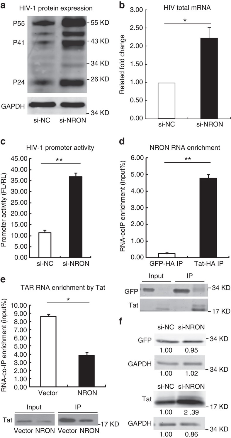
Figure 2. NRON represses HIV-1 transcription by inducing Tat degradation.
(a) HEK293T cells were co-transfected with pNL4-3-deltaE-EGFP provirus plasmid and siRNAs against NRON or nonspecific control. Gag proteins expression was determined by western blotting with anti-p24 antibody at 72 h after transfection. (b) Total HIV-1 mRNAs including spliced and unspliced mRNAs were detected with real-time qRT–PCR in the same HEK293T cells described in a (n=3). (c) HEK293T cells were co-transfected with pHIV-Pro-Luc reporter plasmids, pcDNA3.1-Tat-HA, pRL-CMV (as a transfection normalization reporter) and siRNAs against NRON or nonspecific control. The promoter activity was determined by dual-luciferase reporter assay at 48 h after transfection (n=3). (d) Overexpressed NRON RNA was co-immunoprecipitated by HA-tagged Tat in TZM-bl cells, and the enriched RNA was determined by real-time qRT–PCR. RNA co-immunoprecipitated by HA-tagged GFP was set as a control (n=3), and the precipitated proteins were detected by western blotting. (e) The pcDNA3.1-NRON or empty vector was co-transfected with pcDNA3.1-Tat-HA into TZM-bl cells, the TAR-Luc RNA was enriched by RNA co-immunoprecipitation with anti-HA agarose and quantified with real-time qRT–PCR (n=3). The precipitated proteins were detected by western blotting. (f) Tat and control GFP were detected by western blotting on NRON knockdown in HEK293T cells. Numbers indicated the fold change related to control. Data in b–e show mean±s.d. (error bars). Results in a,f represent three independent experiments. *P<0.05, **P<0.01, Student's unpaired t-test.

