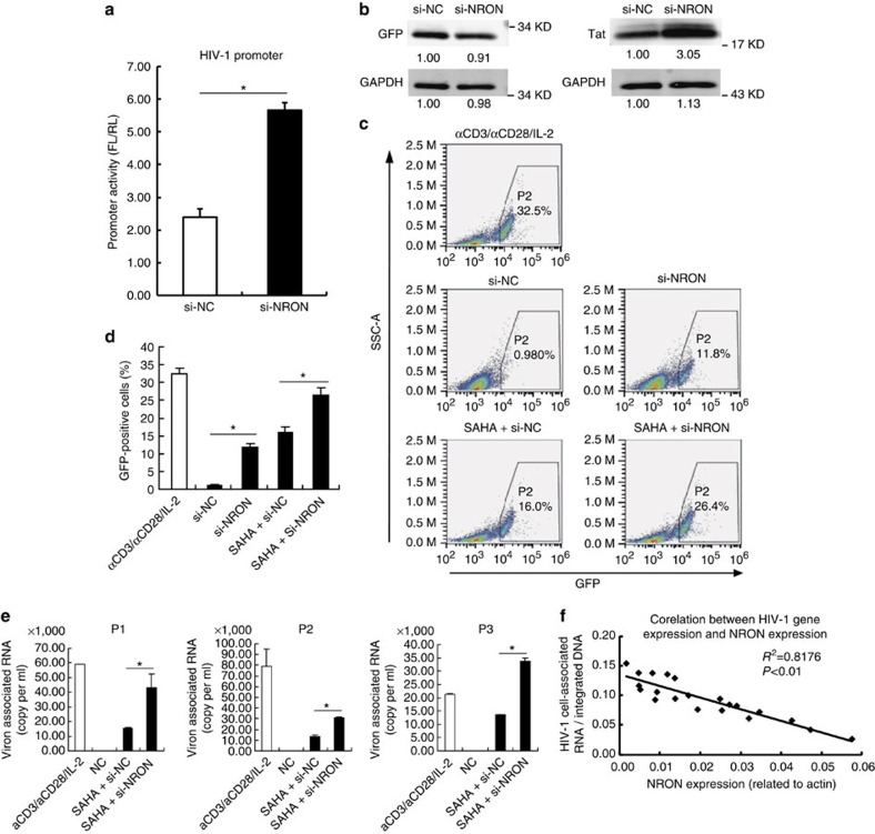
Figure 4. Depletion of NRON reactivates HIV-1 viruses in latently infected CD4+ T lymphocytes.
(a) Primary resting CD4+ T lymphocytes were nucleofected with HIV-1 promoter reporter system plasmids, pcDNA3.1-Tat-HA and siRNAs against NRON or nonspecific control. The promoter activity was determined with dual-luciferase reporter assay at 48 h after transfection (n=3). (b) Tat and control GFP were detected by western blotting on NRON knockdown in nucleofected primary resting CD4+ T lymphocytes. Numbers indicated the fold change related to the control. (c) The latently infected cells were transfected with NRON siRNAs or nonspecific control, or were transfected with siRNAs in combination with the treatment of SAHA, and detected by FACS at 48–72 h post transfection. The GFP+ ratio indicated the reactivation level (d; n=3). (e) Resting CD4+ T lymphocytes isolated from HIV-1-infected individuals on suppressive cART were transfected with siRNAs in combination with the treatment of SAHA. After 48 h, HIV-1 virion-associated RNAs in the supernatants were isolated and detected with real-time qRT–PCR (n=3). (f) The intracellular HIV-1 RNA and NRON RNA expression levels were detected in resting CD4+ T lymphocytes isolated from HIV-1-infected individuals on suppressive cART (n=20), and the correlation between the HIV-1 RNA and NRON RNA levels was shown. The simple linear regression analysis was performed and linear regression line was shown. Data in a,d,e show mean±s.d. (error bars). Results in b represent three independent experiments. *P<0.05, Student's unpaired t-test.

