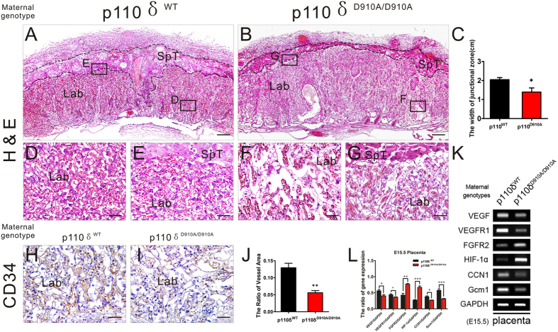
Figure 3. The Defects of Placental Vascularization in p110δ Inactive Female Mice.
Histological appearance of the placenta. (A,B) The representative H&E stained transverse sections of placentas from p110δWT (A) and p110δD910A/D910A (B) female mice at E15.5. (C) The statistical result about the width of junctional zone in placentas from p110δWT (n = 7), and p110δD910A/D910A (n = 8) female mice at E15.5. (D,G) Higher magnification images of labyrinth zone from the sites that indicated by dotted squares in (A,B). (H,I) The immunohistochemical test against CD34 antibody on transverse sections of placentas. (J) The statistical result about the quantity of vessels in the labyrinth zone (K) The RT-PCR data showed the mRNA expression of placental VEGF, VEGFR1, FGFR2, HIF1-α, CCN1 and Gcm1 of E15.5 placenta in p110δWT and p110δD910A/D910A female mice. (L) The statistical result of RT-PCR. The statistical data are expressed as the mean ± S.D. *P < 0.05, **P < 0.01, ***P < 0.001 and ns: P > 0.05. Scale bars: 500 μm (A,B), 50 μm (D–I).

