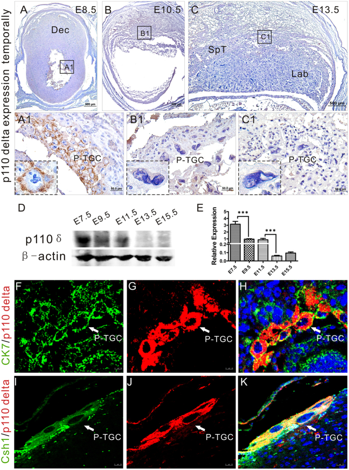
Figure 4. p110δ expresses in P-TGCs in early post-implantation period.
p110δ expresses in P-TGCs. (A–C) Immunohistochemistry against p110δ antibody was performed on transverse sections of tissues from p110δWT female mice at E8.5 (A), E10.5 (B) and E13.5 (C). (A1–C1) Higher magnification images from the sites as indicated by the dotted box in (A–C). (D) Western Blot test at different stages in p110δWT placental biogenesis. (E) The statistical result of western blot data in (D). (F–H) Dual immunofluorescent test against p110δ and CK7 on gestational tissue at E8.5. (I–K) Dual immunofluorescent test against p110δ and Csh1 on gestational tissue at E8.5. Dec, decidua; P-TGC, primary trophoblast giant cell; Csh1, chorionic somatomammotropin hormone 1 (placental lactogen). The statistical data are expressed as the mean ± S.D. ***P < 0.001. Scale bars: 500 μm (A–C), 50 μm (A1–C1), 25 μm (F–K).

