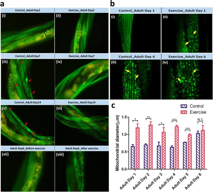
Figure 2. Investigation of the body wall sarcomeres and mitochondria in C. elegans before and after exercise.
(a) Micrographs of body wall sarcomeres at adult days 2, 7, and 14. Sub-images (i–vi) were acquired with a 100× oil immersion objective. The transgenic strain, RW1596 [myo-3p::GFP::myo-3], was used. Sub-images (vii) and (viii) were acquired before and after exercise, respectively. Some of the muscle fibers are indicated by the red arrows. (b) Micrographs of mitochondria changes between the untreated and exercise-treated C. elegans. A transgenic strain, SJ4103[myo-3p::GFP(mit)], was employed. The sub-images (i,ii) and (iii,iv) show that the mitochondrial morphology becomes larger and denser after exercise at adult day 1, however, it compromises at adult day 6. A number of the fused mitochondria are indicated by the yellow arrows. (c) Plot of the mean diameters of mitochondria varying from adult days 1 to 6 (n = 40). The mean diameter of the mitochondrion is larger during the early adult stages in the exercise-treated than the untreated worms. However, this improvement is compromised as the sarcomere integrity downgrades because of age-related degeneration.

