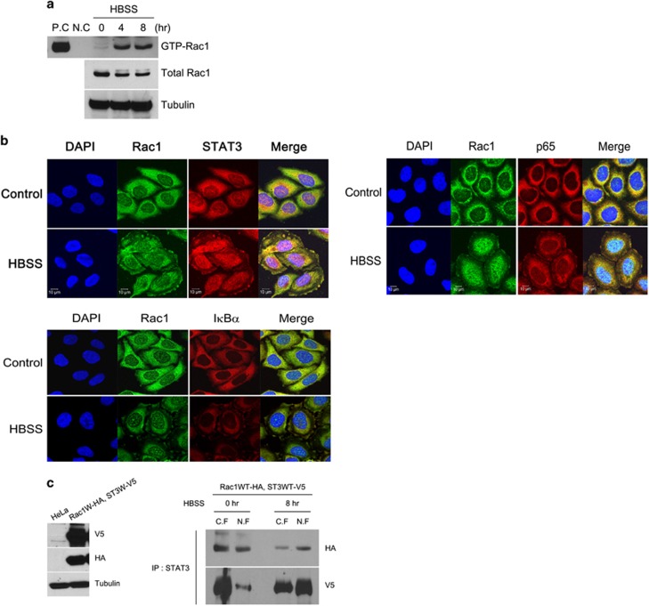
Figure 2.
Rac1 is activated and colocalized with STAT3 or p65 in starved cancer cells. (a) HeLa cells were incubated in HBSS for the indicated time periods, and total cell lysates were incubated with PAK PBD agarose beads. Bound Rac1 was detected with western blotting using a Rac1 Ab (upper panel). The amount of total Rac1 was also measured with western blotting (lower panel). PC, positive control (GTPγS), NC, negative control (GDP). (b) HeLa cells incubated in HBSS for 4 h were fluorescence-stained with anti-Rac1 Ab, anti-STAT3 Ab, anti-p65 Ab and DAPI. (c) HeLa cells expressing vector or Rac1 wild-type HA (Rac1WT-HA) and STAT3 wild-type V5 (ST3WT-V5) were western blotted with V5 Ab, HA Ab and tubulin Ab (left). HeLa cells expressing Rac1WT-HA and ST3WT-V5 were incubated with HBSS for 0 or 8 h. Nuclear and cytoplasmic fractions were prepared, immunoprecipitated with anti-STAT3 Ab and western blotted with anti-HA Ab and anti-V5 Ab (right). DAPI, 4′,6-diamidino-2-phenylindole; HBSS, Hank's balanced salt solution; STAT3, signal transducer and activator of transcription 3.

