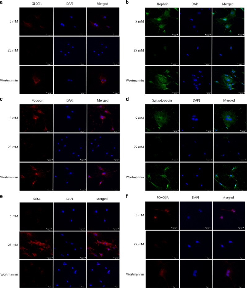
Figure 3.
The localization of glucocorticoid-induced transcript 1 (GLCCI1), podocyte-specific markers and proteins involved in the phosphoinositide 3-kinase (PI3K) signaling pathway in podocytes. (a) In podocytes, GLCCI1 (red, Alexa 568 conjugated) was localized in the 4',6-diamidino-2-phenylindole (DAPI)-stained nuclei. No GLCCI1 localization was observed in the 25 mM D-glucose-treated podocytes. However, GLCCI1 was regulated by wortmannin treatment. (b–d) The podocyte-specific proteins nephrin (green, Alexa 488 conjugated), podocin (red) and synaptopodin (green) showed a reactivity pattern similar to GLCCI1 in podocytes treated with wortmannin. (e) Serum/glucocorticoid-regulated kinase 1 (SGK1; red) was observed only in the 25 mM group. We confirmed that treatment with wortmannin decreased SGK1 expression. (f) Forkhead box O3 (FOXO3A; red) was observed in the 5 mM group. A lower FOXO3A signal was accompanied by high reactivity of SGK1 in the 25 mM group. Localization of FOXO3A was regulated by treatment with wortmannin. Original magnification: × 400. Scale bar, 50 μm.

