Figure 6.
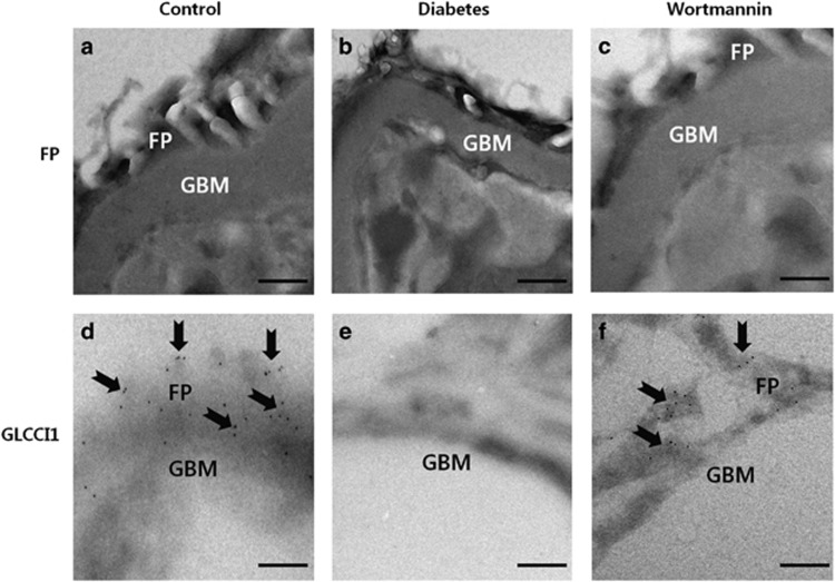
Localization of glucocorticoid-induced transcript 1 (GLCCI1) in podocyte foot processes (FPs) by immunoelectron microscopy. (a) FPs formed by normal podocytes in the control group. (b) Effacement of FPs by podocyte injury in the diabetic group. Podocytes were disrupted by streptozotocin (STZ) treatment. (c) Podocyte FPs were regulated around the glomerular basement membrane (GBM) by treatment with wortmannin. Wortmannin could restore podocytes to their original condition. (d) Immunogold particles (arrowheads) marking GLCCI1 (10 nm gold) were observed in podocyte FPs of the control group. (e) No labeling was observed in podocyte FPs of the diabetic group. (f) Localization of GLCCI1 in podocyte FPs was observed in the wortmannin-treated diabetic group. Scale bar, 200 nm.

