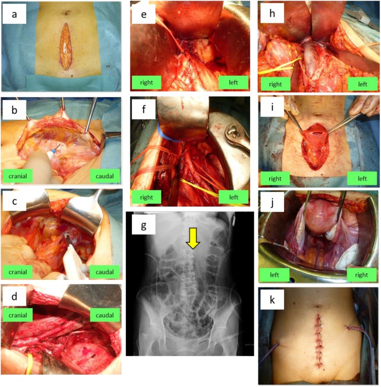
Figure 1.
Abdominal extraperitoneal pelvic and para-aortic lymphadenectomy via the retroperitoneal approach followed by intraperitoneal total abdominal hysterectomy and bilateral salpingo-oophorectomy for endometrial cancer. (a) An approximately 12-cm incision is made in the lower abdomen. (b) The connective tissue space between the rectus abdominis sheath and the parietal peritoneum is developed toward the left inguinal region. (c) After the inferior epigastric vessels and the round ligament are identified, these structures are transected near the pelvic wall. (d) The peritoneal sac containing organs such as the intestinal tract is separated mainly from the external iliac artery and vein, and the pelvic lymph node area is developed. The paravesical space, obturator nerve, ureter (yellow vascular tape), and lateral umbilical ligament (white vascular tape) are identified. The external iliac, external suprainguinal, obturator, internal iliac, common iliac nodes and presacral nodes are dissected in this order. (e) The left transversalis fascia is separated, and the peritoneal sac containing the intraperitoneal organs is freed. Using an Octopus Retractor with a long hook, the sac is displaced from the common iliac artery in the cranial direction along the left side of the aorta to develop the para-aortic area. The inferior mesenteric artery (IMA) (red vascular tape) is identified. (f) The para-aortic lymph nodes to the left side, anterior and posterior of the aorta are dissected in the order of nodes caudal to the IMA followed by nodes cranial to the IMA. (g) Dissection of para-aortic lymph nodes up to the level of L2 is confirmed by the position of the vascular clip on a postoperative plain abdominal X-ray film (arrow). (h) Para-aortic lymph nodes to the right of the inferior vena cava and nodes lying between the vena cava and aorta are dissected in the order of nodes caudal to the IMA followed by nodes cranial to the IMA. (i) After lymphadenectomy is completed, a midline incision is made in the peritoneum to enter the peritoneal cavity. (j) The uterus and bilateral adnexae are dissected via the extrafascial method. (k) The midline peritoneal incision is closed with sutures. Then a retroperitoneal drain is placed, the wound is closed, and surgery is completed.

