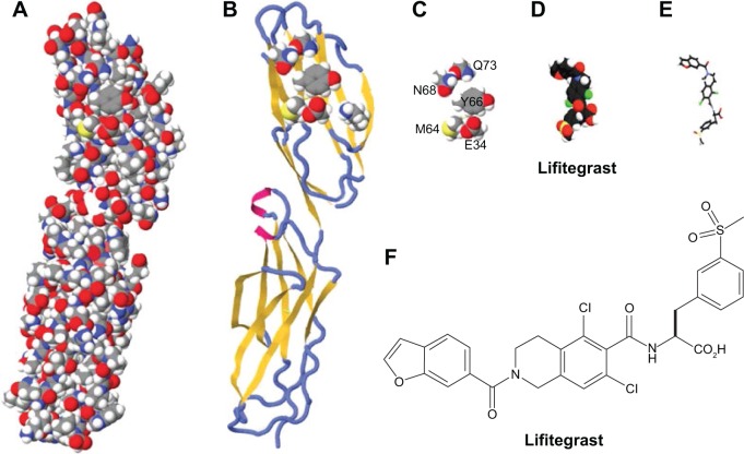
Figure 2.
The evolution of the lifitegrast design targeting LFA-1.
Notes: (A) The space-filling model of the first two domains of ICAM-1 as determined by X-ray crystallography; (B) ribbon diagram of the first two domains of ICAM-1 with the superimposed binding residues of E34, K39, M64, Y66, N68, and Q73 in domain 1; (C) space-filling depiction of the six amino acid side chain binding residues identified by alanine point mutagenesis of the ICAM-1 epitope that binds to LFA-1; (D) the molecular structure of lifitegrast represented using a space-filling model; (E) the three-dimensional structural form of lifitegrast as represented from image D; (F) the structural formula of lifitegrast.
Abbreviations: ICAM-1, intercellular adhesion molecule-1; LFA-1, lymphocyte function-associated antigen-1.

