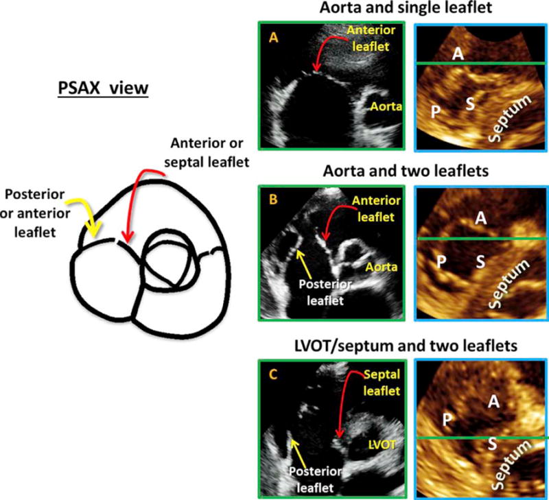
Figure 5.

In the PSAX view (left) the leaflet is closest to the aorta is always the anterior or septal and never the posterior. Near the RV free wall, the posterior or anterior leaflet could be seen but never the septal leaflet. The anterior leaflet was depicted if a single leaflet was seen on the 2D image (A with corresponding MPR result to the right). The A-P combination was noted if two leaflets were seen with a central coaptation point together with the aortic valve (B with corresponding MPR result to the right). When the 2D plane intersected below the aortic valve, in the area of the left ventricular outflow tract or septum, and the aortic valve was not seen throughout the cardiac cycle, the leaflets imaged were the septal and the posterior leaflet (C with corresponding MPR result to the right).
