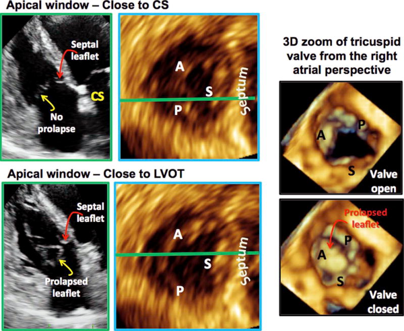
Figure 9.

Using the proposed nonstandard TV views to localize TV prolapse. In the A4C view with coronary sinus (left, top row), the prolapse was not seen, suggesting that the prolapse did not involve the posterior or the septal leaflets. In the A4C view with left ventricular outflow tract (LVOT), the prolapse was seen (left, bottom row), suggesting that the pathology was related to the anterior leaflet. MPR analysis in each of the 2D views confirmed these conclusions (left, top and bottom MPR views). Three-dimensional zoom of the TV as seen from the right atrial perspective also confirmed these findings. CS, Coronary sinus.
