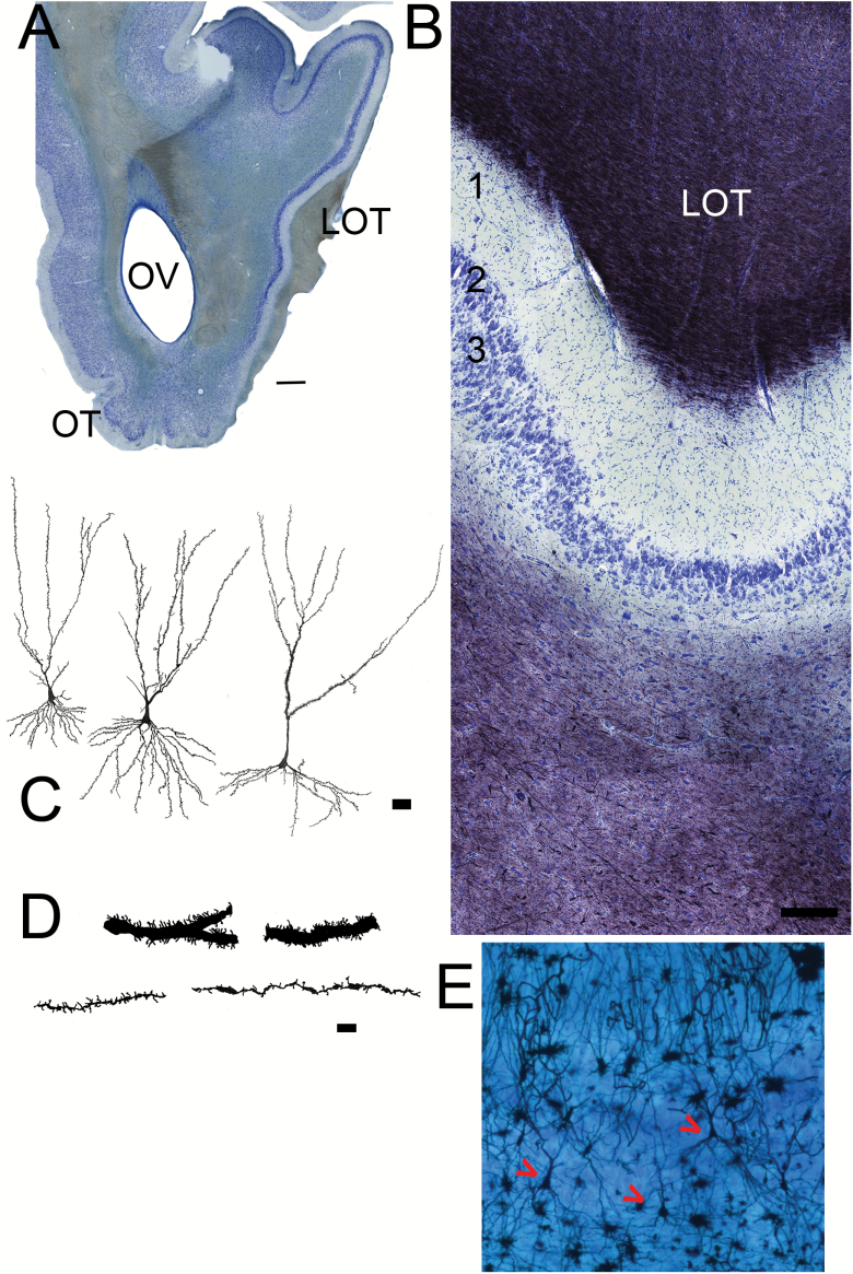
Figure 7.
Sections from the anterior PC. (A) Low magnification picture (scale bar = 1mm). The olfactory tubercle has emerged on the ventral side of the olfactory peduncle. The LOT is visible on the right (lateral) side. The APC occupies the entire lateral side of the peduncle. (B) Nissl (blue)/myelin (black) staining. Superficial at top. As in Figure 5, the top of the section contains myelinated fibers and collaterals from the LOT that thin toward the deeper regions indicating the beginning the Layer 1 (1A) of the APC. Under the cell poor Layer 1 are cellular layers 2 and 3. Scale bar = 200 µm. (C) Camera lucida drawings of pyramidal neurons from the APC. Scale bar = 50 µm. (D) Higher magnification drawings of sections of apical dendrites from APC pyramidal neurons. Some cells have large caliber dendrites with many spines (top), others have much finer processes with fewer spines (bottom). Scale bar = 10 µm. (E) Photomicrograph of Golgi/Nissl section showing three pyramidal neurons (red arrows).

