Figure 5.
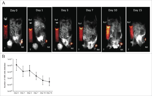
19F-labeled NK cell migration in NSG mice bearing human GD2+ melanoma and hu14.18-IL-2 treatment. 10 × 106 19F-labeled (4 mg/mL PFPE for 24 h) human NK cells were injected on the left flank subcutaneously into NSG mice (n = 2) bearing human melanoma (M21) tumor on the right flank (T: Tumor). On days 0–2 and 7–9, 50 µg/50 µL hu.14.18-IL-2 immunocytokine was injected i.t. On days 0 and 7, 1 × 10⁶ IU/0.2 mL rh-IL-2 was injected i.p. (A) Mice were imaged for 1H and 19F by MRI for 42 min at different time points. Here, a T2-weighted 1H image was acquired for enhanced tumor visualization. Composite 19F/1H images are depicted for 1 mouse. (B) Illustrates the number of NK cells remaining at the site of injection at each time point for both mice. Day 0 refers to the day of implantation of 19F-labeled human NK cells. Values represent the mean ± SEM of one single experiment, n = 2.

