
Figure 6. Histological analysis shows degeneration of the photoreceptors in mutant mice.
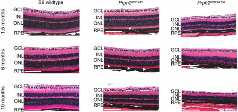
Wild-type, heterozygous and homozygous mutant retinas are shown at the indicated time-points. Note the shortened outer segments (OS) in the mutants (arrow). Displaced nuclei are frequently seen in the OS of heterozygous mutants at 6 and 10 months (arrowhead). A marked reduction of the outer nuclear layer (ONL) in the heterozygote and its absence in the homozygous mutants is observed at 10 months. Scale bar indicates 100mm.