
Figure 8. Histological analysis shows similarities in the degeneration of the photoreceptor between Prph2nmf193/+ and Nr2e3rd7/rd7 mutants.
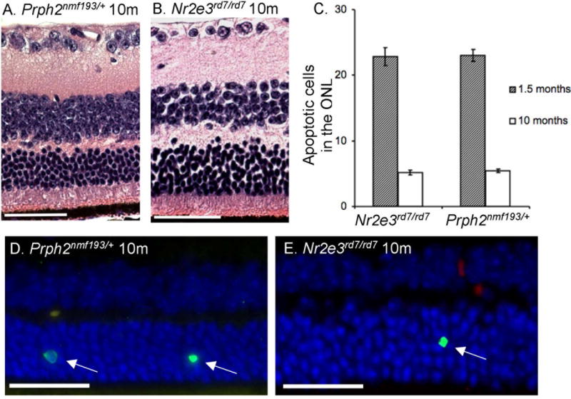
Hematoxylin and eosin staining shows outer nuclear layers reduced to 5–7 cells thick in the central retina of both mutants (panels A, B). Scale bar indicates 50 mm. TUNEL staining was used to identify apoptotic nuclei in the ONL per 5mm section in both mutants at 1.5 and 10 months (panels C–E). Wild-type controls showed negligible TUNEL staining at both time points. A total of 100 microns of sections were counted per mouse.