
Figure 9. Similar degeneration observed in photoreceptor degeneration in Prph2nmf193/+ and Nr2e3rd7/rd7 mutants, and direct targeting of Prph2 by Nr2e3.
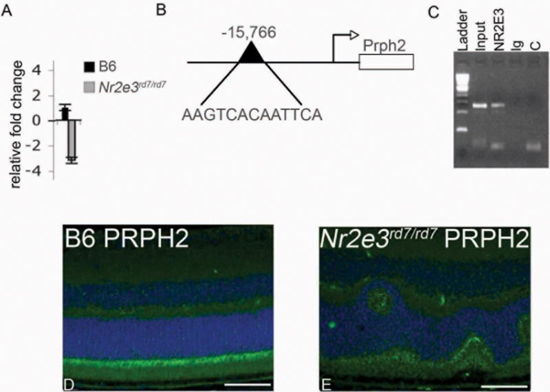
A. Quantitative real time PCR analysis showing 3.2 fold reduced expression of Prph2 in P21 Nr2e3rd7/rd7 relative to control. B. Schematic representation of Prph2 genomic sequence indicating location and sequence of NR2E3 binding response element (RE). C. Chromatin immunoprecipitation assay showing direct binding of NR2E3 to Prph2 RE sequence. L-ladder, I-input (no Antibody, positive control); NR2E3 (NR2E3 antibody precipitated sample); Ig (IgG antibody precipitated, negative control); C (no template negative control). D (B6 control), E (Nr2e3rd7/rd7) immunohistochemisty of P30 retina sections. Photoreceptor outer segments labeled with PRPH2 (green) and DAPI (blue nuclear stain) show reduced expression in Nr2e3rd7/rd7 retinas.