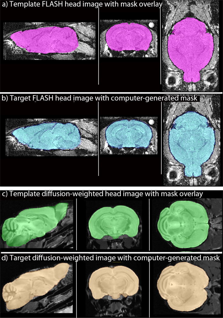
Fig. 3. Examples of application of computer-stripping to two other imaging modalities.

(a and b) 3D MR images acquired with a FLASH protocol are equally well stripped by automation, as shown in selected sagittal, coronal and axial slices: (a) Template whole head image with its hand-drawn mask overlay, extracted brain shown in pink. (b) Target image and its computer-generated mask, created from the hand-drawn mask in (a) overlaid on a different image from the same cohort of mice imaged with the same imaging parameters. Extracted brain shown in blue. (c and d) Slices from 3D images acquired with a diffusion-weighted protocol (Bearer et al., 2009): (c) Template image with its hand-drawn mask used to generate the mask for the target image in (d). Extracted brain in green. (d) Target image with computer-generated mask. Extracted brain in copper.
