
Figure 1.
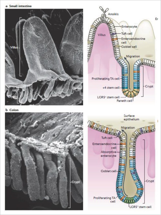
Intestinal epithelium: Scanning electron micrograph (SEM) (left) of (a) small intestine, villus can be observed, (b) colon which has no villi but multiple crypts. Diagram of the intestine (right) showing the crypt which contain stem cells which divide into proliferating transint-amplyfing (TA) cells which differentiate into cells such as enterocytes, goblet cells (which secrete mucus) and tuft cells. +4 stem cells are believed to act as reserve stem cells to replace LGR5+ stem cells during injury thus restoring the normal cell renewal process. During cell renewal, which takes 3-5 days, cells migrate from the crypt up toward the villi. At the top of the villi, anoikis or programmed cell death occurs. This is also the site of cell sloughing during intestinal injury. Reproduced with permission.46