
Figure 4.
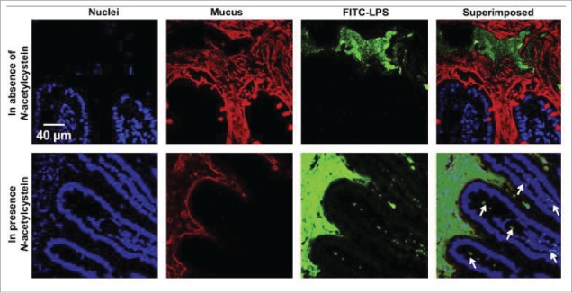
Confocal images showing the intestinal absorption of FITC-LPS (green) after its oral administration in the absence/presence of a mucolytic agent (N-acetylcysteine). In the presence of the mucolytic agent, the mucus layer (red) became thinner, and FITC-LPS was observed underneath the epithelium (indicated in the superimposed image by the white arrows), an indication of the intestinal absorption of LPS. Reproduced with permission.62