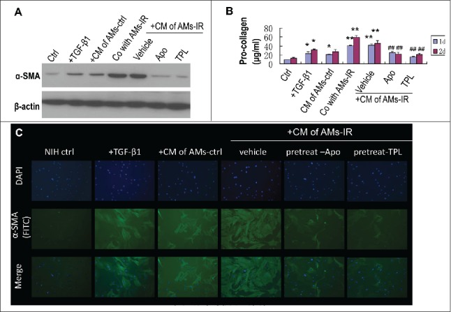
Figure 3.

TPL and Apocynin abrogated AMs-IR induced myofibroblast activation and pro-collagen secretion. NIH3T3 cultured with conditioned medium (CM) of AMs for 1 or 2days, or NIH3T3 co-cultured with AMs-IR using transwell for 1 or 2days. (A) The level of α-SMA in myofibroblasts was measured by Western blot. (B) The level of pro-collagen in CM was assayed by Sirus red method (n = 3). (C) NIH3T3 cells were grown on glass coverslips and cultured with CM for 2days, and then the α-SMA expression in myofibroblasts was detected by immunofluorescent staining(× 400). +TGF-β1: NIH3T3 treated with TGF-β1 (5 ng/ml); + CM of AMs-ctrl: AMs-ctrl culture alone for 1day, then the CM of AMs-ctrl was transferred to the culture well of NIH3T3; + CM of AMs-IR (vehicle, Apo or TPL): AMs-IR treated without (vehicle) or with Apocynin (Apo, 300 mM) or TPL (5ng/ml) for 1day, then the CM was transferred to the culture well of NIH3T3; Co with AMs-IR: transwell® co-culture of AMs-IR with NIH3T3. AMs-IR were cultured in transwell inserts (0.4 μm pore size), and NIH3T3 cells were cultured in the bottom of transwell plate. ** P < 0.01 vs. ctrl; ## P < 0.01 vs. CM of AMs-IR (vehicle).
