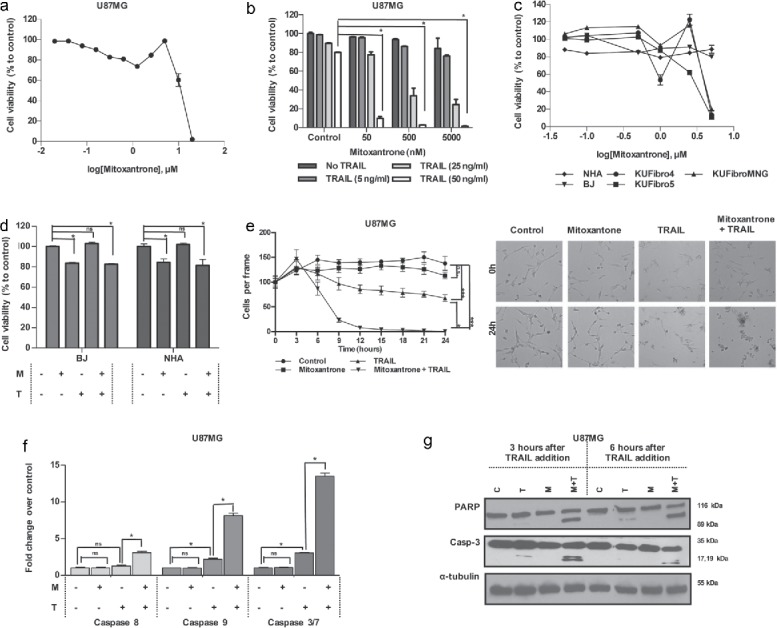
Figure 3.

Effect of a DNA damaging agent, Mitoxantrone, on GBM and non-malignant cell viability (A) Viability of U87MG cells upon 0.02–20 μM of Mitoxantrone treatment. (B) The effect of different dose combinations of Mitoxantrone and TRAIL on U87MG cell viability. (C) The effects of Mitoxantrone doses between 50–5000 nM on primary human fibroblasts: KUFibro5, KUFibroMNG and established cell lines, BJ and human astrocytes (NHA). (D) The effect of the combination Mitoxantrone (500 nM) and TRAIL 50ng/ml) on BJ and NHA. (E) Live cell imaging of U87MG cells treated with Mitoxantrone, TRAIL and the combination for 24 hours. Left: graph depicting viable cell numbers, Right: representative snapshot images at t = 0 and t = 24 hours. Statistics were performed by 2-way ANOVA with Tukey's post hoc testing. (F) Caspase-8, −9 and −3/7 level activity of Mitoxantrone and/or TRAIL-treated U87MG cells. (G) Detection of apoptosis markers, cleaved PARP and cleaved caspase-3 protein levels, by Western Blot. *Indicates p <0.05, *** indicates p<0.0001 and ns stands for nonsignificant.
