
Figure 1.
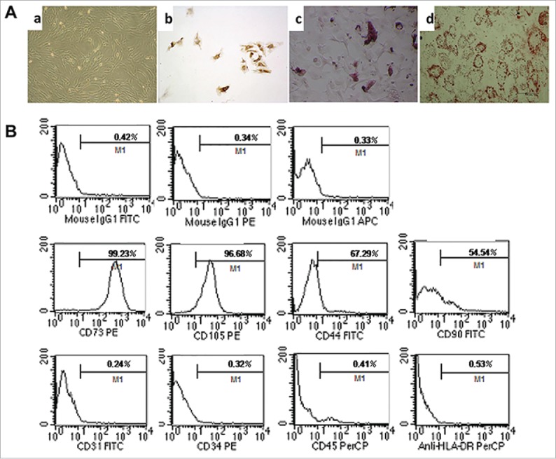
Characteristics and differentiation potential of MSCs derived from umbilicalcord tissues. (A) Morphology of UC-MSCs Magnification:×100. After chondrogenic differentiation conditions, MSCs differentiate into chondrogenic-like cells and immunohistochemically stained positive for type II Collagen; ×100. After osteogenic-specific induction, the MSCs were stained with Alizarin red; ×100. After inducing adipogenic differentiation, the cells showed many small lipid vacuoles, as assessed by Oil Red O staining;×100. (B) Flow cytometric analysis showing the MSC cells surface antigens: positive for mesenchymal lineage markers (CD44, CD73, CD90 and CD105), negative for haematopoietic and endothelial markers (CD31, CD34 and CD45), and negative for HLA-DR.