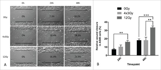
Figure 5.
Wound healing assay of A549 cells 3 d after exposure to AIR and FIR. (A) Representative wound-healing images taken at 0h, 24h and 48h with respective wound-closure ratios. Magnification x100. (B) Wound-closure analysis after AIR and FIR normalized to 0% at 0h. Experiments were repeated 3 times. Bars represent standard deviation from one representative experiment.

