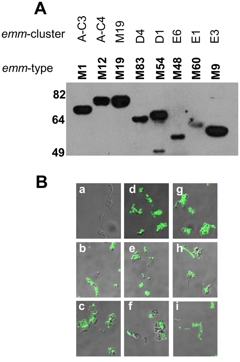
Fig 4. Anti-SV1 antibody binding to recombinant M-proteins and GAS surface.
(A) M-proteins were electrophoresed in 12% SDS-PAGE gels transferred to nitrocellulose membrane and probed anti-SV1 antisera. The emm-type and emm-cluster assignment are shown at the top of the figure. (B) Immunofluorescent microscopy demonstrating that anti-SV1 antibodies bind to the surface of multiple GAS emm-types. The nine strains are (a) JRS145 (emm-negative), (b) 5448, (c) PRS9, (d) PL1, (e) PRS30, (f) PRS42, (g) PRS15, (h), PRS20 and (i) PRS55. Images are shown as overlays of bright field and fluorescent images. No fluorescence was observed when PBS sera was used in the same assays (data not shown).

