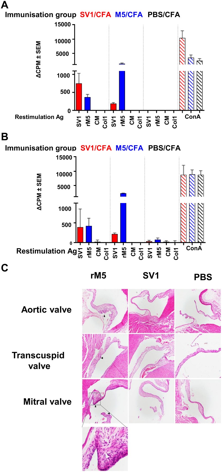
Fig 5. T cells from SV1 immunized rats do not cross-react with tissue antigens and do not cause inflammatory changes in cardiac tissue.
(A) Data is presented as the change in counts per minute (ΔCPM) between stimulated and unstimulated groups (A) T-cells responses inSV1/CFA, M5/CFA or PBS/CFA immunized rats following restimulation with SV1, M5, cardiac myosin or collagen. (B) T cell responses in SV1/alum, M5/alum or PBS/alum immunized rats following restimulation with SV1, M5, cardiac myosin or collagen. (C) Representative histology sections of Lewis rat cardiac tissues (x100 magnification) following immunization with recombinant M5-protein, SV1 or PBS with Freund’s adjuvant. Areas of inflammation are indicated with a black arrow. A higher magnification image (x400) of an inflamed mitral valve from a rat immunized with M5 is shown. Mice that received PBS only or SV1 as antigen had no evidence of inflammatory changes in cardiac tissue.

