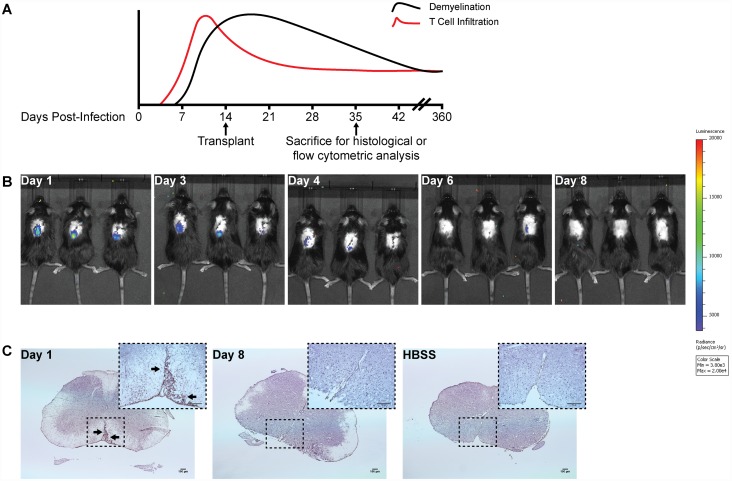
Fig 2. Human iPSC-derived NPCs are rapidly rejected following intraspinal transplantation in JHMV-infected mice.
(A) Timeline highlighting the relationship of T cell infiltration and demyelination in response to JHMV infection of the CNS as well as when cells are transplanted into the spinal cord and when animals are sacrificed to assess histology and immune cell infiltration into the CNS. (B) In vivo bioluminescence imaging revealed EB-NPCs could be detected in the spinal cords of transplanted animals as early as day 1 post-transplant (p.t.) and were undetectable by day 8 pt. (C) Representative brightfield images of coronal spinal cord sections from EB-NPC transplanted mice stained with SC121, a monoclonal antibody specific for human cytoplasm. Arrows indicate SC121+ regions. Human cells were detected in ventral white matter regions at day 1 p.t. but could be not be detected by day 8 p.t., confirming rejection of EB-NPCs. Scale bars = 100 μm.

