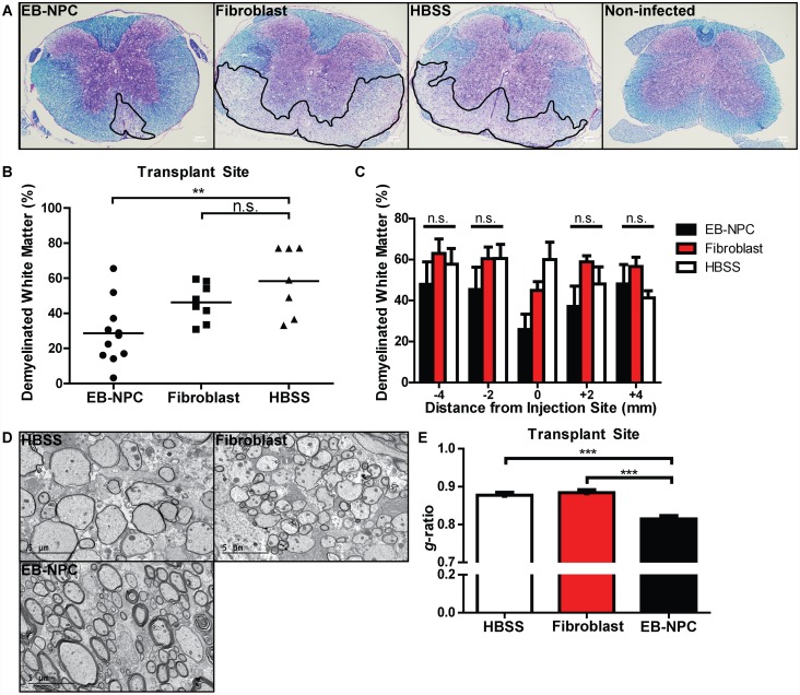
Fig 4. Focal remyelination in animals transplanted with EB-NPCs.
(A) Representative brightfield images of coronal spinal cord sections stained with luxol fast blue (LFB) and counter-stained with hemotoxylin and eosin (H&E). (B) Quantification of demyelination in the ventral white matter of EB-NPC, fibroblast, and HBSS injected mice revealed significantly (p < 0.01) reduced demyelination at the injection site in the spinal cords of EB-NPC-transplanted mice. (C) Quantification of demyelination in areas adjacent to the injection site revealed that reduced demyelination was not sustained along the rostrocaudal axis. (D) Representative electron micrographs of coronal spinal cord sections from HBSS, fibroblast, and EB-NPC-injected mice. (E) Analysis of the ratio of the axon diameter vs. total fiber diameter (g-ratio) confirmed enhanced remyelination at the transplant site of EB-NPC-injected mice compared to controls (p < 0.001). For (B) and (C), data represents two independent experiments with n = 11 (EB-NPC), n = 8 (Fibroblast), and n = 7 (HBSS) animals per group. For (D), ≥ 300 axons were measured per experimental group. All data is presented as average ± SEM and was analyzed using one-way ANOVA followed by Tukey’s multiple comparison test.

