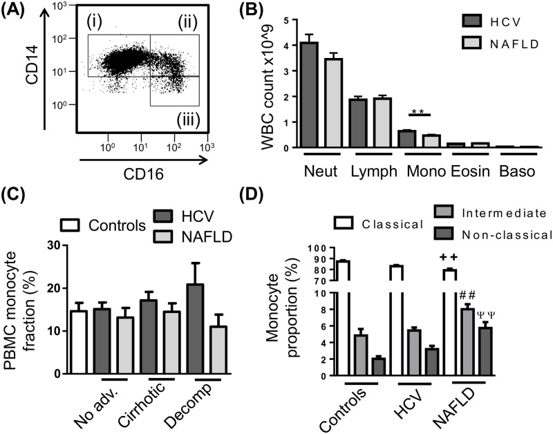
Fig 1. Peripheral blood monocyte subset distribution in chronic liver disease (CLD).
(A) Representative flow cytometry plot of the 3 main monocyte subsets, i—CD14highCD16- “Classical”, ii—CD14highCD16+ “intermediate”, iii—CD14+CD16+ “non-classical” monocytes. (B) Distribution of peripheral leukocyte lineages obtained from patient haematology full blood counts. (C) Proportion of monocytes in isolated peripheral blood mononuclear cell (PBMC) in control subjects and CLD patients with no-advanced fibrosis (No adv.), cirrhosis or decompensated cirrhosis (Decomp). (D) Distribution of monocyte subsets in the PBMC fraction of control subjects and CLD patients. (Data represented as mean +SEM, ** ++ ## ψψ P<0.01; D, significance shown vs corresponding control subset; Neut, neutrophils; Lymph, lymphocytes; Mono, monocytes; Eosin, eosinophils; Baso, basophils)

