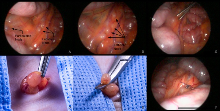
Fig 1.
A. Laparoscopic view of a paracolonic and two left-colic nodes draining a portion of the descending colon. B. Four left colic nodes draining a different portion of the descending colon. C. Use of the Maryland forceps to grasp the mesentery adjacent to a left-colic node prior to externalization. D. Externalized node held with a rat tooth forceps. E. Externalized node clamped with a mosquito hemostat prior to ligation. F. 4–0 PDS at ligature site in the abdomen.

